Cells: The smallest units of life
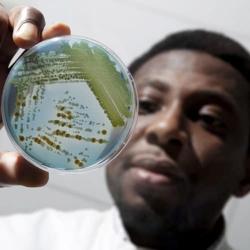
All living organisms are made up of cells. Organisms that have just one cell, such as bacteria, are called unicellular organisms. Multicellular organisms, on the other hand, can have several billion cells.
Cells are the basic units that make up all the tissues in an organism, such as the skin, muscles and neurons in the human body, the leaves on a tree, or the petals on a flower.
What characterizes cells?
Cells are the basic units of life. Without exception, all living organisms have at least one cell.
Each cell is autonomous and can carry out the vital functions necessary to its survival, such as nutrient uptake, adaptation and reproduction.
Every cell is surrounded by a plasma membrane that separates and protects it from the outside environment. Cells contain a gelatinous liquid called cytoplasm as well as different organelles. The vast majority of cells have a complete set of their organism’s DNA.
Cells (and the living organisms they form) can be put into two main categories depending on whether they lack or contain a nucleus: prokaryotes and eukaryotes.
Prokaryotic cells
The word “prokaryote” is derived from Greek and means before the nucleus (pro = before, karyon = kernel/nucleus).
Prokaryotic cells are the cells of unicellular organisms, such as bacteria and archaea. Their distinctive feature is that they lack a nucleus surrounded by a nuclear membrane. The DNA of prokaryotes floats in the cell cytoplasm and bundles together into a less structured region called the nucleoid.
Prokaryotic cells can only produce offspring through asexual reproduction, which means by a single parent without the formation and fusion of sex cells. Unlike sexual reproduction, which combines genetic material from two parents, asexual reproduction creates new individuals that are genetically identical to the parent, i.e., clones.
Eukaryotic cells
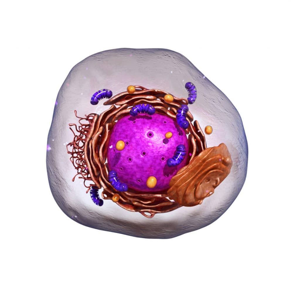
The term “eukaryote” means true nucleus in Greek (eu = true, karyon = kernel/nucleus). These cells have a nuclear membrane that encloses the genetic material, along with structures called organelles that perform different functions. These cells are larger and more complex than prokaryotic cells and generally form multicellular organisms.
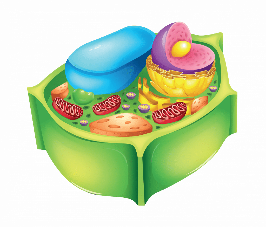
There are two main types of eukaryotic cells: animal cells and plant cells. The tables below outline their similarities and differences.
Structures shared by all eukaryotic cells
| Cell structure | Definition | Location |
| Nucleus | The nucleus is the largest organelle in eukaryotic cells. It is surrounded by a nuclear membrane and contains almost all of the organism’s DNA, as well as the elements to decode and replicate DNA. | Inside the cell |
| Cytoplasm | This part of the cell contains a fluid rich in proteins (cytosol), as well as the cell’s organelles. Many chemical reactions take place in the cytoplasm. | Inside the cell |
| Plasma membrane | This membrane surrounds the cell and plays a role in communication and exchanges between the inside and the outside of the cell. | Separates the cell from its environment |
| Rough endoplasmic reticulum (RER) | The site where proteins are processed. RER is found just outside the nuclear membrane. It is called “rough” because, unlike smooth endoplasmic reticulum, it contains ribosomes. | Cytoplasm |
| Smooth endoplasmic reticulum (SER) | SER synthesizes lipids, stores substances and eliminates molecules that are harmful to the cell. SER is said to be smooth because, unlike the RER, it lacks ribosomes. | Cytoplasm |
| Ribosomes | The site of protein synthesis. | Rough endoplasmic reticulum and cytoplasm |
| Cytoskeleton | The cytoskeleton acts as a scaffold to provide structural support and maintain the cell’s shape. It is responsible for cell movements and the movement of material within the cell. It helps regulate cellular processes. | Cytoplasm |
| Mitochondria | These organelles convert food into energy that is usable by the cell (ATP). | Cytoplasm |
| Vesicles | These structures transport cellular material and proteins within the cell and toward the outside of the cell. | Cytoplasm |
| Golgi body | This organelle processes and sorts proteins. | Cytoplasm |
Animal cells
| Cell structure | Definition | Location |
| Lysosomes | These membrane sacs are filled with enzymes that “digest” different molecules. They break down or recycle things the cell no longer needs, such as parts of organelles and damaged proteins. They also play a role in the immune system and help destroy harmful invaders in the body. | Inside the cell |
Plant cells
| Cell structure | Definition | Location |
| Chloroplasts | These light-sensitive organelles contain a pigment called chlorophyll. They transform carbon dioxide into sugar using the sun’s energy in a process called photosynthesis. | Inside the cell |
| Cell wall | The cell wall is an additional protective layer that withstands the environment’s physical and chemical stressors. | Separates the inner cell from its outer environment |
| Vacuoles | These compartments have multiple functions. They act as reservoirs to store nutrients, water and pigments and play a role in detoxification. They also provide structural support by exerting pressure on the cell wall and interacting with other organelles to recycle cellular components. The different types of vacuoles are what help plant cells thrive in a variety of environments. | Inside the cell |
Cells: Protein assembly lines!
Cells can be described as protein kitchens. Their nucleus contains DNA, which is like a recipe book containing instructions on how to create proteins and which, like a chef, relays those instructions to the kitchen—or cell—team. Organelles work like specialized line cooks on different tasks of the protein recipe.
Not every part of the genome is expressed in every cell. Only the proteins needed for the cell to function are expressed. There are different types of cells for all the different tissues of an organism, each with their own roles. These cells include neurons, muscle cells, skin cells, gland cells and many more. Cells also form all the tissues in plants, fungi, animals and all other living organisms.
- Neurons are very elongated and have dendrites that make a huge number of connections with other cells so that they can pass on information very quickly. They must produce a lot of proteins for this special cellular architecture (cytoskeletal proteins).
- The epithelial cells of our intestine have microvilli, which are finger-like structures that enlarge the surface area to absorb nutrients. These cells produce proteins that let them absorb nutrients (transmembrane proteins) and secrete immunoglobulins, which are proteins of the immune system.
- Goblet cells are elongated and secrete mucus. They produce the proteins in mucus, which include mucin and antibacterial enzymes.
- Eggs are the largest cells in the human body, while sperm cells are the smallest. These cells have proteins on their surface that let them bind during fertilization.
- Muscle cells contract to let the body move. These cells need to produce lots of myosin, which are the proteins that help these cells transform energy into movement.
Cell cycle
The cell cycle is the life cycle of a cell. It has two phases: the interphase and the mitotic phase, also known as the cell division phase.
Why do cells divide?
Like all living things, cells eventually die. The remaining cells must divide to replace the missing cells. This is how organisms grow and also heal from their wounds.
The interphase is the period when the cell plays its role in the organism and grows. This phase is also when it duplicates its chromosomes for cell division.
The mitotic phase of cell division produces two child cells that are genetically identical to the parent cell.
The DNA is duplicated, then distributed to each side of the parent cell (mitosis). The cell then divides into two child cells (cytokinesis).
- Prophase: The chromosomes condense into tightly packed coils. The mitotic spindle, a structure composed of protein fibres, begins to form in the cell nucleus.
- Metaphase: The chromosomes align into a structure called the equatorial plate. The mitotic spindle fibres attach to the centre of the chromosomes.
- Anaphase: Each of the sibling chromatids is pulled to opposite ends of the parent cell by mitotic spindle fibres. The cell stretches out to draw the genetic material to each side.
- Telophase: The sibling chromatids reach the opposite poles and decondense into their interphase shape. New nuclei begin to form, and the cell prepares to completely separate in two.
- Cytokinesis: The cytoplasm and cell membrane divide into two child cells.

